
Alopecia
Introduction
This chapter provides an overview of alopecia (excluding the genetic
hair shaft defects). Differentiating between scarring and non-scarring alopecia
is particularly important because scarring alopecia is irreversible and so
early intervention is required in order to try and minimise hair loss.
This chapter is set out as follows:
Aetiology
The hair cycle
- Hair follicles undergo a
repetitive sequence of growth and rest known as the hair cycle. To
understand the different causes of hair loss it is important to have a
comprehension of the hair cycle:
- Anagen - is the period of
active growth and may last for several years. Under normal circumstances
80-90% of hair follicles on the human scalp are in anagen at any one time
- Catagen - at the end of
anagen, epithelial cell division declines and ceases, the hair follicle
enters an involutionary phase known as catagen
- Telogen - the period between
the completion of follicular regression and the onset of the next anagen
phase is known as telogen, during which time the hair is shed
- Kenogen - in the human scalp,
hair follicles may remain in a state of latency, also known as kenogen,
for a prolonged period after the hair is shed
- The term telogen
effluvium is broadly used to describe any cause of diffuse
hair shedding in the telogen phase, and results from the
premature termination of anagen with a subsequent reduction in the length
of the hair cycle
Clinical findings
Understanding the
normal scalp
- Dermoscopy is being increasingly used
to help diagnose the type of alopecia, and consequently reduce the need
for scalp biopsies, which in themselves may not be diagnostic
- In the normal scalp several
hairs can be seen in each follicular unit, all with a similar width. There
is often mild loose and non-perifollicular scale
A logical approach
to alopecia
1. Is it non-scarring
or scarring?
2. If non-scarring is
it male or female pattern alopecia?
3. If non-scarring is
the hair loss diffuse?
4. If non-scarring is
it more patchy?
5. Scarring alopecia -
is it one of the cicatricial (circular / oval shaped patches of alopecia)
causes or is it something else?
1. Non-scarring or
scarring alopecia?
The first step to making a diagnosis is to differentiate whether the
alopecia is non-scarring or scarring:
- Non-scarring alopecia
- No clinically visible
inflammation is noted in most cases
- Atrophy absent
- Tufting absent
- Tends to have preserved
follicular openings
- Scarring alopecia (figure
19)
- Clinical inflammation is
frequently, but not always, present
- Loss of follicular openings
- Atrophic, often with a
shiny appearance
- Variable dermoscopic
features, which may include follicular heads having a black 'clubbed'
appearance
- 'Dolls hair' tufting -
mainly seen in folliculitis decalvans, and occasionally in other causes
of long-term severe scarring
2. Non-scarring
alopecia - male and female pattern
- Male pattern alopecia (figures 1-2)
- Recession of the frontal
hair line, mainly in a triangular pattern is the characteristic finding,
later followed by thinning of the vertex
- Dermoscopic features - in
affected areas there will be fewer hairs in each follicular unit and more
than 20% of the hairs will be small and fine ie miniaturised
- Refer to the related
chapter Alopecia - male and female pattern alopecia for
more information
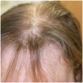
- Female pattern alopecia (figure 3)
- The Ludwig pattern - is
characterised by a diffuse thinning of the centroparietal region with
maintaining of the frontal hair line. It is the most common type in
women, occasionally also observed in men
- The Christmas tree pattern
- this is similar to the Ludwig pattern in that the Christmas tree
pattern shows diffuse centro-parietal thinning, but additionally, the
frontal hair line is breached
- Dermoscopic features - as
above
- Refer to the related
chapter Alopecia - male and female pattern alopecia for
more information
- If associated with
hirsutism or other signs of virilisation also refer to the related
chapter Hyperandrogenism
3. Non-scarring
alopecia - diffuse
Acute:
- Drugs (acute and/or chronic)
- In particular chemotherapy agents, which cause hairs to fall out in their anagen growth phase, referred to as anagen effluvium (figure 4)
- Acute telogen effluvium
- Normally occurs two to
three months after a triggering event such as pyrexia
(in particular a body temperature of 102.5 degrees Fahrenheit and above),
child-bearing, major surgery, weight loss and certain medications
- In one-third of cases no
trigger is identified
- Unless the trigger is
repeated, spontaneous complete regrowth occurs within three to six
months
More gradual:
- Other
causes of telogen effluvium
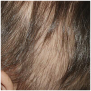
- Thyroid disorders (figures
5-6)
- Profound iron deficiency
anaemia (figure 7) can cause a diffuse hair loss. The
association between iron deficiency and no or mild anaemia and chronic
diffuse hair loss is controversial, however, a trial of iron supplements
is worthwhile for ferritin levels < 40
- Increasing age
- Many drugs have
been implicated with hair loss in the telogen phase although good
evidence for a link is not often evident. A dose-related diffuse alopecia
is commonly seen with acitretin
- Crash dieting
- Various - disorders causing malabsorption,
hepatic and renal disorders, malignancy, SLE and dermatomyositis can
all cause telogen hair loss
- Zinc deficiency, both acquired (from
long-standing parenteral nutrition) and inherited, can be associated with
a severe telogen effluvium. Acrodermatitis enteropathica is
a rare genetic disorder associated with the malabsorption of zinc and
characterised by diarrhoea, an inflammatory rash around the mouth and /
or anus, and alopecia
- Alopecia areata: occasionally the
initial hair loss may be diffuse
- Secondary syphilis (figure
8):
is non-inflammatory and can present with a diffuse pattern of alopecia, a
moth-eaten pattern or a combination of both. The scalp is the most
commonly affected area. However, the eyebrows, eyelashes, axilla, pubis,
chest, and legs can also be affected. Alopecia syphilitica often
accompanies other mucocutaneous symptoms of secondary syphilis, but it
can be the only presenting symptom
- Investigations - all patients
with diffuse alopecia require a FBC, ferritin and TFT. The need for any
additional tests depends on the history / examination as above
- Chronic telogen effluvium (CTE)
- CTE is a primary idiopathic
and often self-limiting condition affecting middle-aged women
- Patients, often with longer
and thick hair, describe large amounts (often 50%) of hair loss and
the patient may bring the hair to the consultation
- On examination the patient
has a good head of hair appropriate to their age with no widening
of the central parting line
- Despite of the shortened
hair cycle, which may go on for several years, women with CTE should be
reassured that the natural history is NOT that of a
progressive alopecia (unless the patient also has female pattern
alopecia, the two conditions can co-exist)
- There is no treatment other
than reassurance
- Occasionally such patients
may have a dysmorphophobia, a psychiatric condition, the
hallmark of which is a fixation on an imaginary flaw in the patient's
physical appearance, or in cases in which a minor defect truly exists the
patient exhibits an inordinate amount of anguish
- Provide a patient information leaflet
4. Non-scarring
alopecia - more patchy
- Alopecia areata (figure 9)
- The characteristic
initial lesion is a circumscribed, totally bald, smooth patch.
The skin within the bald patch is normal or slightly reddened. Short
broken hairs (exclamation mark hairs) are often seen around the margins
of active expanding patches of alopecia
- Dermoscopic features - yellow dots
and also black dots if a hair is broken off at the root, coiled ‘pig
tail’ hairs (not specific but most commonly seen in alopecia areata),
exclamation mark hairs
- Refer to the related
chapter Alopecia areata for more
information
- Tinea capitis (figure 10)
- Can cause non-scarring or
scarring alopecia
- Hair loss tends to be
patchy, but is often associated with subtle inflammation / scale
- Dermoscopic features - scale, broken
hairs with black dots, comma-shaped hairs
- Refer to the related
chapter Tinea capitis for more
information
- Trichotillosis (formerly called
trichotillomania) - hair pulling (figures 11-15)
- Trichotillosis is a
behavioural disorder characterised by compulsive hair pulling associated
with an increase in tension prior to the pulling and a sense of relief
when the hair is pulled out
- It is more common in women
and can occur in people from all walks of life
- The hair manipulations
frequently occur in patients engaged in sedentary activities
- Distribution
- The patches may be single
or multiple
- The frontal and temporal
scalp are most commonly affected, but any site can be involved
- The degree of involvement
may vary from only a few square centimeters to large areas of scalp
- Unlike in alopecia areata
- The patches of alopecia
may take on unusual shapes
- It is uncommon for the
hair to be completely lost within a patch of alopecia, and the remaining
hair is firmly anchored in the scalp
- Dermoscopic featues - hair
length is not uniform. Other features can include newly growing short
hairs with tapered ends, broken short terminal hairs, vellus or
indeterminate hairs, black dots, empty follicular orifices
- The establishment of a good
relationship between the physician and patient or parents of the affected
child is important, as is the making of a confident diagnosis
- Management is mainly
psychological support. Attempts should also be made to try and identify
triggers for the patient's behaviour
- Traction alopecia (figures 16-17)
- Is due to certain hair
styles which cause a sustained pull on the hair
- The pattern of hair loss is
often distinctive and reflects the direction of traction
- Traction alopecia is most
commonly seen in Afro-Caribbean hair styles when the hair is tightly
braided. The hair loss commonly begins in the temporal regions and in
front of / above the ears
- The presence of retained
hairs along the frontal and / or temporal rim, termed the “fringe sign,”
is a common finding in both early and late traction alopecia, and is a
useful clinical marker of the condition
- Can be associated with
headaches
- Changes are reversible if
the style is changed early - if braids continue to be used their
directions should be alternated, and must be loose
- If there is no history of
significant traction the term primary marginal alopecia is
sometimes used
5. Scarring
alopecia
- Infection
- Bacterial infection eg a
severe staphylococcal infection
- Tinea - refer to the
related chapter Tinea capitis for more
information (figures 19-20)
- Cicatricial alopecia - circular-oval shaped
patches of scarring
- Lichen planopilaris (figure 21)
- Follicular erythema and
keratotic plugs, which are commonly located at the periphery of
expanding areas of alopecia
- Dermoscopic features - perifollicular
scale and inflammation
- Refer to the related
chapter Lichen planus - follicular lichen planus for
more information
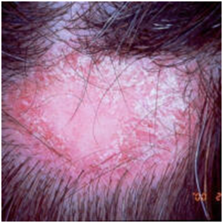
- Discoid lupus erythematosus - DLE (figure 22)
- Much more common in women
- Scarring alopecia occurs
in 20% of men and 50% of women with DLE
- Although DLE often
presents on the face, the scalp can be the only affected site
- The scalp is often itchy
- Affected patches are
erythematous and scaly with follicular plugging
- Dermoscopic features - often chaotic with
yellow rough looking follicular plugs, telangiectasia and white or dark
areas. Erythema and scale may be more pronounced around the periphery of
areas of alopecia
- Patients are treated along
the lines of cutaneous lupus with UV-protection, super-potent topical
steroids, intralesional steroids and antimalarial therapy
(hydroxychloroquine +/- mepacrine). Other treatments sometimes used
include retinoids (studies undertaken used acitretin), dapsone,
thalidomide and occasionally immunosuppressive therapies (refer to the
related chapter on Lupus erythematosus for more
information)
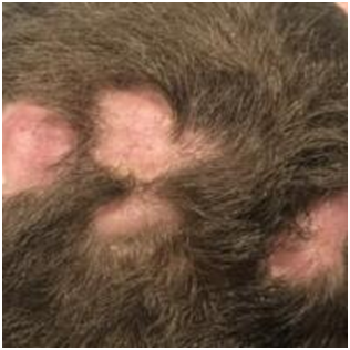
- Folliculitis decalvans (figure 23)
- Patients usually present
with one or more round patches of scarring alopecia usually surrounded
by pustules, crusting and sometimes erosions
- Nearly always starts on
the crown and expands outwards
- Dermoscopic features - marked 'dolls
hair' tufting, scale, perifollicular and interfollicular erythema,
pustules. Crusting will be present if the inflammation is very active
- Refer to the related
chapter Folliculitis decalvans for more
information
- Central centrifugal cicatricial alopecia CCCA
(figure 24)
- Is a condition that
predominantly affects women of African ethnicity
- It usually presents in the
fourth decade with a female to male ratio of approximately 3:1
- The aetiology is unknown
although it is probably genetic in some
- Traction (from heated
styling instruments or chemical straighteners), pattern hair loss and
iron-deficiency may co-exist. CCCA is also associated with
hirsutism
- Clinically there is a diffuse
scarring alopecia that begins at the crown and spreads forwards.
The alopecia is incomplete with a number of hairs remaining within the
area of scarring. There tends to be little or no erythema
- Dermoscopic features - in dark skin the
normal scalp appearance is a pigment network with regular white dots. In
CCCA there are white-grey halos around follicles and subtle irregular
white dots
- Mycology to rule out tinea
capitis is recommended
- No ideal treatment
currently exists. Minimal hair grooming is recommended, but
many patients find this difficult. If there are signs of inflammation
(clinically or histologically) the use of a potent topical or
intralesional corticosteroid may arrest/slow progression. Occasionally
pustules are seen in which case a systemic tetracycline such as
doxycycline or lymecycline may be of value. Other treatments
occasionally used in the presence of inflammation include those used for
lichen planopilaris
- Pseudopelade of Brocq (figure 25)
- Asymptomatic, slowly
progressive, patchy cicatricial alopecia with no evidence
of inflammation
- Two aetiological theories
exist
- It
is primarily an atrophic condition
- It
is the end point of inflammatory conditions such as follicular lichen
planus
- There is no known
effective treatment
Other causes of
scarring alopecia
- Frontal fibrosing alopecia (figure 26)
- A type of lichen planus
- Most common in
post-menopausal women but can affect other age groups and rarely men
- Hair loss first affects the
arms and legs, sometimes years before the eyebrows and then the
scalp
- In the scalp it presents as
a progressive symmetric band-like alopecia, affecting the frontal hair
line, the pre-auricular scalp, and less commonly all the way around the
hairline
- Dermoscopic features -
follicular prominence with hyperkeratosis (excess scaling) around
hair follicles, inflammation can be subtle or absent
- Refer to the related
chapter Lichen planus - follicular lichen planus for
more information
- Acne keloidalis nuchae
- Acne keloidalis nuchae
(AKN) is a condition characterised by follicular-based papules and
pustules that form hypertrophic or keloid-like scars. AKN typically
occurs on the occipital scalp and posterior neck and develops almost
exclusively in young, African-American men. Refer to the related chapter Acne Keloidalis Nuchae for more
information
- Dissecting cellulitis (figure 27)
- More common in black
patients and between the ages of 18-40
- A variant of acne
conglobata
- Presents with painful
nodules and abscesses that taken on a cerebriform appearance. Can become
extensive across the scalp
- Early intervention can
prevent scarring
- Treatment - patients should be referred
urgently, refer to the chapter Dissecting cellulitis
- Pemphigus (figure 28)
- Pemphigus vulgaris affects middle-aged
patients causing mucosal lesions, flaccid blisters on the skin and
erosive scalp changes
- Pemphigus foliaceus is less severe than
pemphigus vulgaris and mucosal lesions are uncommon
- Cicatricial pemphigoid
- Mainly affects elderly
patients. This bullous disorder involves the eyes and / or other mucosal
surfaces causing painful erosions and scarring. Patients with cutaneous
involvement present with tense blisters and erosions, often on the head
and the neck, with scalp involvement in 10% of cases
- Erosive pustular dermatosis of the scalp
(figures 29-31)
- Affects the elderly with
varying degrees of scarring associated with yellow-brown crusts,
pustules, lakes of pus, erosions and ulceration. Previous UV damage,
cryotherapy or other physical insults to the skin appear to play a role.
Treatment is with super-potent topical steroids and UV protection
- Linear morphoea 'en coup de sabre' (figure 32)
- An atrophic band-like area
of alopecia on the frontoparietal scalp and forehead
- Follicular mucinosis (figures 33-34)
- Benign follicular
mucinosis: sometimes referred to as alopecia mucinosa -
a benign condition. Early signs of the disease are the presence of
grouped follicular papules arising in patches / plaques, usually 2-5 cm
in diameter. Hair loss is common from the affected follicles. In some
patients the condition resolves spontaneously and the hair regrows. In
more severe disease complete follicular destruction prevents normal hair
growth. Other features can include acneiform lesions
- Folliculotropic mycosis
fungoides: this
is due to a cutaneous t-cell lymphoma, and is characterised by a
more generalised chronic form in a slightly older age group, with larger
and more numerous plaques on the scalp, face and also the extremities
Images
Please refer to notes on image rights at bottom of the page with regards
to individual image ownership.


2

3

4

5
6

7

8

9

10

11
12

13

14

15

16

17

18

19

20

21
22

23

24

25

26
27

28

29

30

31

32

33

34
Investigations
Investigations depend on the clinical context but can include:
- Swabs of pustules for C
& S - the contents of the pustule should be expressed
- If tinea is suspected,
scrapings of scale and plucked hairs should be sent for mycology
- Non-scarring alopecia can be
multifactorial so consider the following blood tests:
- Diffuse alopecia /
female-pattern alopecia / undetermined alopecia - FBC, ferritin, U&E,
LFT, TFT, zinc levels, and vitamin D. The need for any additional
tests depends on the history / examination eg ANA / ENA for suspected
lupus, VDRL for suspected syphilis
- Female patients presenting with alopecia
and hirsutism / irregular periods or other signs of virilisation need
further investigations - refer to the related chapter Hyperandrogenism
- Biopsies
- Scalp biopsies are not
always needed and in some case do not provide a definitive
diagnosis. The main reason to biopsy is for cases of diagnostic
uncertainty, especially if the outcome may affect management
- If a scalp biopsy is to be
performed it is worth checking with a local pathologist as to what is
required - some departments prefer two 4 mm punch biopsies, one each
for horizontal and vertical sectioning. Biopsies should be
taken from active sites (as opposed to scarred sites)
and angled at the same direction as the hair to try and
get the whole hair follicle. If considering lupus erythematosus a sample
should also be sent for direct immunofluorescence
- In general the decision as
to whether or not a biopsy is needed is best left to a specialist, who
will also be in a better place to correlate the histology result and
clinical findings should a biopsy be performed
Management
- Management depends on the
clinical context - refer to the clinical findings and
investigations above, and related chapters (top right)
- Who to refer
- Patients with scarring
alopeica require an urgent referral - once
scarred the alopecia is irreversible
- Patients with non-scarring
alopecia may also need referring as follows:
- Diagnostic uncertainty
- Some cases of alopecia
areata
- Management issues eg
trichotillomania
- If associated with
moderate-severe emotional distress
- If a wig is required -
wigs can be used as effective ways to cope with alopecia. Local
dermatology departments may be the best contact in terms of obtaining /
finding out where to obtain wigs on the NHS. Patients are given a choice
of monofilament acrylic wigs or synthetic acrylic wigs. Human hair wigs
are normally reserved for patients found on patch tests to be allergic
to acrylic wigs
Assessment Form - use ai mode
Hair & Scalp Health Assessment (Form)
Instructions:
Identify Hair Type (No Score): First, identify your basic hair type for context.
Calculate Your Score: Check all boxes in the Scorecard that apply to you and add up the points.
Read Interpretation: Use your total score to find the corresponding interpretation and recommended actions.
Part 1: Identify Your Hair Type (For Context - No Score)
Type 1 (Straight): Generally straight, from fine/oily to coarse.
Type 2 (Wavy): "S" shaped waves, can be fine, frizzy, or coarse.
Type 3 (Curly): Defined "S" spirals or springy corkscrews.
Type 4 (Kinky/Coily): Tightly coiled, fragile "S" or "Z" pattern.
Part 2: Hair Loss & Scalp Concern Scorecard
Part 3: Score Interpretation
Score: 1-4 (Low Concern)
Interpretation: Your symptoms are mild and may be related to normal hair cycles, styling habits (breakage), or minor stress.
Recommendation: Monitor the situation. Focus on a healthy diet, gentle hair care, and stress management.
Score: 5-9 (Moderate Concern)
Interpretation: A noticeable pattern of hair loss or shedding is present. This is beyond typical daily shedding and warrants attention.
Recommendation: Take Action. Consider over-the-counter Medicine. It is advisable to consult a primary care doctor or dermatologist to identify potential underlying causes (like vitamin deficiencies or hormonal changes).
Score: 10+ (High Concern)
Interpretation: The combination and severity of your symptoms (e.g., rapid loss, scalp inflammation, distinct patterns) strongly suggest an underlying medical condition.
Recommendation: Seek Professional Advice Urgently. Schedule an appointment with a dermatologist. Early and accurate diagnosis is critical to effective treatment and preventing further loss.
Disclaimer: This chart is an informational tool and not a substitute for professional medical diagnosis. Please consult a healthcare provider for any health concerns.


0 Comments